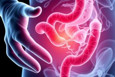
Appendicitis is one of the most common medical emergencies encountered worldwide, especially in young adults and children. While the diagnosis may sometimes seem straightforward, the presentation can vary significantly, making clinical examination essential. One of the key physical signs that physicians rely on is Rovsing’s sign. This clinical sign is a subtle yet important clue that often points toward appendicitis, especially in ambiguous cases. In this article, we will dive deep into Rovsing’s sign in appendicitis — what it is, how it is tested, why it matters, and how it fits into the broader context of diagnosing this painful and potentially dangerous condition.
What Is Rovsing’s Sign?
Rovsing’s sign is a classic physical examination finding used to help diagnose appendicitis. Specifically, it refers to the occurrence of pain in the lower right abdomen when the lower left side of the abdomen is palpated or pressed. Essentially, an examiner applies pressure to the left lower quadrant, and if this maneuver elicits referred pain to the right lower quadrant (where the appendix is typically located), Rovsing’s sign is considered positive.
This sign was first described by Niels Thorkild Rovsing, a Danish surgeon, in the early 20th century. It’s fundamentally based on the anatomical and physiological connections within the peritoneum—the lining of the abdominal cavity—and how irritation involving the appendix causes pain to radiate.
Why Is Rovsing’s Sign Important in Appendicitis Diagnosis?
Appendicitis, an inflammation of the appendix, classically presents with pain that begins around the belly button (periumbilical area) and localizes to the right lower quadrant over time. However, not all patients have textbook symptoms. Especially in children, elderly patients, or pregnant women, the signs can be misleading. Rovsing’s sign becomes a part of what doctors call the physical exam “toolkit,” helping to confirm the suspicion of appendicitis when other symptoms and signs might be equivocal.
It’s important to note that while imaging techniques such as ultrasound and computed tomography (CT) scans have revolutionized appendicitis diagnosis, physical findings like Rovsing’s sign remain valuable. They are quick, cost-free, and can guide the urgency and focus of further testing.
The Physiology Behind Rovsing’s Sign
To grasp why pressure on the left lower quadrant causes right lower quadrant pain, consider the innate sensitivity of the peritoneum. The peritoneal lining is highly sensitive, and inflammation in one part can cause pain when adjacent or distant parts are manipulated. When the left side of the colon is pressed, the movement of gas and content through the intestines may result in distension or stretching that aggravates the inflamed appendix on the right. This leads to localized pain referred to that area.
How to Test Rovsing’s Sign: A Step-by-Step Guide
Performing a Rovsing’s sign test requires a gentle but deliberate technique, typically done by a healthcare professional during a physical exam.
- Step 1: The patient lies supine (on their back) with relaxed abdominal muscles.
- Step 2: The examiner places their hand on the left lower quadrant of the abdomen, just above the pelvic bone.
- Step 3: Gradually and gently, pressure is applied to this area.
- Step 4: The examiner observes whether this causes pain or tenderness in the right lower quadrant.
If the patient experiences sharp or localized pain in the lower right abdomen during this maneuver, Rovsing’s sign is considered positive. The presence of this sign raises the likelihood of appendicitis but, like all clinical signs, it is not definitive on its own.
Other Related Physical Signs in Appendicitis
Rovsing’s sign frequently forms part of a larger collection of physical tests used to diagnose appendicitis. These include:
| Physical Sign | Description | Indication |
|---|---|---|
| McBurney’s Point Tenderness | Tenderness over McBurney’s point, located between the anterior superior iliac spine and the umbilicus on the right side. | Classic sign indicating localized inflammation of the appendix. |
| Psoas Sign | Pain elicited by passive extension or active flexion of the right hip. | Suggests irritation of the psoas muscle by an inflamed retrocecal appendix. |
| Obturator Sign | Pain elicited by internally rotating the flexed right hip. | Indicates irritation of the obturator internus muscle near the appendix. |
| Rebound Tenderness | Pain felt upon rapid release of pressure on the abdomen. | Signals peritoneal irritation. |
Combining Rovsing’s sign with these other signs enhances diagnostic accuracy, guiding timely management.
Interpreting Rovsing’s Sign: Limitations and Diagnostic Accuracy

No physical sign is perfect, and Rovsing’s sign is no exception. Numerous studies have attempted to quantify its sensitivity and specificity. Generally, Rovsing’s sign has moderate sensitivity and relatively low specificity, meaning that while its presence increases suspicion for appendicitis, its absence does not rule it out, nor does its presence guarantee the diagnosis.
Some key points on interpreting Rovsing’s sign include:
- Positive Rovsing’s sign: Enhances suspicion for appendicitis, especially when accompanied by other signs and symptoms.
- Negative Rovsing’s sign: Does not exclude appendicitis. Many patients with appendicitis may not exhibit this sign.
- False positives: Can occur in other conditions causing peritoneal irritation, such as diverticulitis or pelvic inflammatory disease.
Thus, Rovsing’s sign should be considered as part of a comprehensive clinical evaluation including history, laboratory tests (like white blood cell count), and imaging when necessary.
Combining Rovsing’s Sign with Other Diagnostic Tools
In modern medicine, clinicians rarely rely solely on physical signs. The diagnosis of appendicitis involves a blend of clinical scores, laboratory data, and imaging. Here’s how Rovsing’s sign integrates:
Clinical Scoring Systems
Several scoring systems incorporate physical signs, including Rovsing’s sign, to estimate the likelihood of appendicitis. One widely used example is the Alvarado score:
| Parameter | Points |
|---|---|
| Migration of pain to the right lower quadrant | 1 |
| Anorexia (loss of appetite) | 1 |
| Nausea or vomiting | 1 |
| Tenderness in the right lower quadrant | 2 |
| Rebound pain | 1 |
| Elevated temperature (>37.3°C) | 1 |
| Leukocytosis (increased white blood cells) | 2 |
| Shift to the left (increased neutrophils) | 1 |
Note that Rovsing’s sign specifically is incorporated in some variations or considered part of the right lower quadrant tenderness assessment, reflecting the relevance of appendiceal irritation.
Laboratory Tests
Elevated white blood cell count and inflammatory markers, like C-reactive protein, support the diagnosis in conjunction with clinical signs. However, laboratory findings are non-specific and cannot replace a thorough physical examination.
Imaging Studies
Ultrasound and CT scans provide more definitive evidence of appendiceal inflammation. The physical exam, including Rovsing’s sign, often determines whether such imaging is necessary urgently.
Conditions That Can Mimic Rovsing’s Sign in Appendicitis
A positive Rovsing’s sign doesn’t always mean appendicitis. Other conditions causing irritation or inflammation of the peritoneum on the right side can produce similar findings. These mimics include:
- Diverticulitis: Particularly sigmoid diverticulitis on the left side can cause generalized peritoneal irritation.
- Pelvic Inflammatory Disease: Infection of the female reproductive organs may cause pelvic tenderness and refer pain.
- Kidney Stones: Can present with flank pain and sometimes overlap with lower quadrant abdominal pain.
- Mesenteric adenitis: Inflammation of the lymph nodes in the abdomen, especially in children, may mimic appendicitis.
- Gastrointestinal infections: Many infections cause generalized abdominal pain and discomfort that may confuse clinical findings.
This highlights the importance of combining Rovsing’s sign with the broader clinical picture for accurate diagnosis.
Historical and Modern Perspectives on Rovsing’s Sign

Since its discovery, Rovsing’s sign has remained a popular physical exam technique, taught to medical students as part of a classic surgical exam. In the pre-imaging era, such signs were critical to determine the need for surgical intervention. Today, while imaging can confirm diagnosis with high accuracy, expert clinicians still value their role in rapid bedside assessment.
Recent research continues to assess the diagnostic value of Rovsing’s sign and related physical examinations, often noting variability depending on examiner skill and patient population. Some studies encourage combining multiple signs for higher diagnostic confidence.
Tips for Clinicians on Using Rovsing’s Sign Effectively
- Ensure the patient’s abdominal muscles are relaxed to reduce false positives from muscle guarding.
- Communicate clearly with patients during the exam to differentiate between superficial tenderness and deeper pain.
- Use gentle, incremental pressure to avoid causing unnecessary discomfort.
- Always interpret Rovsing’s sign within the context of the patient’s entire clinical presentation.
The Patient Experience: What Does It Feel Like?
Patients might wonder what pain triggered by Rovsing’s sign indicates. The discomfort caused by pressing on the left lower abdomen and feeling pain on the right can be disconcerting. Often, this pain is sharp, localized, and prompts attention to the seriousness of their condition. Understanding that this referred pain occurs due to inflammation can help patients appreciate the clinical examination process and the urgency of their symptoms.
Summary Table: Rovsing’s Sign in Appendicitis
| Aspect | Details |
|---|---|
| Definition | Pain in right lower quadrant upon palpation of left lower quadrant. |
| First Described | Early 20th century by Niels Thorkild Rovsing. |
| Physiological Basis | Referred pain due to peritoneal irritation and intestinal movement. |
| How Tested | Gentle pressure on left lower abdomen causes right-sided pain. |
| Clinical Use | Helps support diagnosis of appendicitis alongside other findings. |
| Limitations | Not definitive; moderate sensitivity; can be positive in other conditions. |
| Other Signs to Use Together | McBurney’s tenderness, psoas sign, obturator sign, rebound tenderness. |
Conclusion
Rovsing’s sign represents a fascinating and practical aspect of the physical exam in appendicitis, revealing how careful clinical observation can provide critical diagnostic information. Though modern imaging has enhanced diagnostic precision, this simple bedside test remains a valuable tool for healthcare providers. Understanding Rovsing’s sign — from its physiological basis to how it is tested and interpreted — helps both clinicians and patients appreciate the complex nature of appendicitis diagnosis. Ultimately, the sign underscores the timeless importance of blending clinical skills with modern technology to ensure timely, effective treatment for patients with right lower quadrant pain.