Ultrasound for appendicitis diagnosis is an essential medical imaging technique that doctors frequently turn to when a patient presents symptoms of acute abdominal pain. It’s a non-invasive, safe, and effective way to evaluate the appendix and help detect inflammation, thereby confirming or ruling out appendicitis. But what makes ultrasound the go-to choice in many cases? And how does it compare to other diagnostic tools? In this comprehensive guide, we will walk you through everything you need to know about the role of ultrasound in diagnosing appendicitis, including its benefits, limitations, and practical use in clinical settings.
Understanding appendicitis—the inflammation of the appendix—is crucial because it’s a common cause of sudden abdominal pain that often requires urgent surgical removal. Misdiagnosis or delay can lead to complications like a ruptured appendix and serious infections. Hence, timely and accurate diagnosis is paramount. Ultrasound, especially in pediatric and pregnant patients, serves as a frontline imaging technique, offering a quick and effective snapshot of the appendix without the risks associated with radiation.
How Does Ultrasound Work in Diagnosing Appendicitis?

Ultrasound uses high-frequency sound waves to create images of the inside of the body. When it comes to appendicitis diagnosis, a specially trained technician or radiologist applies a transducer to the patient’s lower right abdomen, where the appendix is located. The sound waves bounce off tissues and organs and are converted into live images on a monitor. This allows doctors to check for signs of an inflamed appendix, such as enlargement, thickening of the wall, or associated fluid collections.
One of the fundamental reasons ultrasound is valued in appendicitis diagnosis is its real-time imaging capability. The technician can gently press the transducer to see if the patient experiences pain—a clinical sign known as “sonographic tenderness.” This tangible interaction helps correlate the ultrasound findings with physical symptoms, which can be crucial in ambiguous cases.
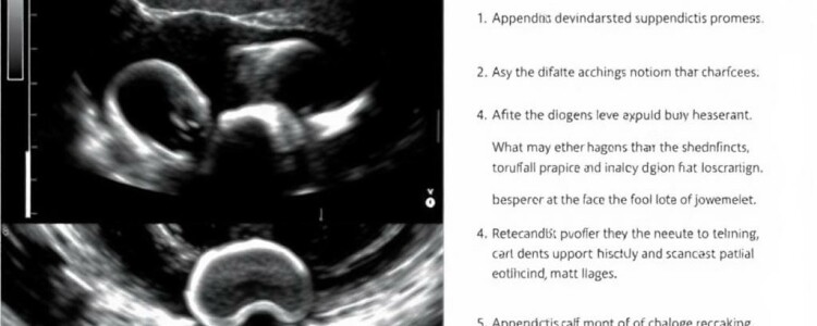
Key Ultrasound Findings in Appendicitis Diagnosis
A normal appendix may be hard to spot on ultrasound because it is small and can blend in with surrounding intestines. However, an inflamed appendix shows several tell-tale features that radiologists look for:
- Enlarged appendix diameter greater than 6 mm
- Non-compressibility under the probe’s pressure
- Wall thickening and increased echogenicity of surrounding fat
- Presence of an appendicolith (calcified deposit inside the appendix)
- Fluid collections or abscess near the appendix
Doctors often combine these ultrasound findings with clinical symptoms such as fever, rebound tenderness, and elevated white blood cell counts for a more accurate diagnosis.
Advantages of Using Ultrasound for Appendicitis Diagnosis
Ultrasound stands out among diagnostic tools for several reasons, particularly when compared with CT scans or MRI:
| Advantage | Description |
|---|---|
| Safety | Ultrasound uses no ionizing radiation, making it ideal for children, pregnant women, and repeated follow-ups. |
| Non-invasive | The procedure is painless and involves no needles or injections, often requiring only a few minutes. |
| Availability | Ultrasound machines are widely available in emergency departments and clinics worldwide. |
| Cost-effective | Compared to CT or MRI, ultrasound is less expensive and more accessible. |
| Real-time imaging | Allows dynamic evaluation, such as observing compressibility and tenderness responses. |
These advantages help explain why ultrasound is often the first-line imaging technique for suspected appendicitis, particularly when radiation exposure needs to be minimized.
Ultrasound vs. Other Imaging Modalities for Appendicitis Diagnosis
While ultrasound offers many benefits, it is important to understand its place relative to other diagnostic tools:
- CT Scan: Generally considered the gold standard with high accuracy, CT scans provide detailed cross-sectional images but expose patients to radiation, which can be risky, especially for younger individuals.
- MRI: Increasingly used, particularly in pregnant patients, MRI offers excellent soft tissue contrast without radiation. However, it is more expensive, time-consuming, and less readily available in emergency settings.
- Physical Examination & Lab Tests: While vital for initial assessment, these alone cannot conclusively diagnose appendicitis without imaging support.
In many healthcare settings, ultrasound serves as the initial test, and if results are inconclusive, a CT scan or MRI might follow. This stepwise diagnostic approach balances accuracy, safety, and cost.
Challenges and Limitations of Ultrasound for Appendicitis Diagnosis

Despite its clear advantages, ultrasound is not without challenges when used for appendicitis diagnosis.
Operator Dependence
One of the major limitations is the reliance on the skill and experience of the sonographer and interpreting radiologist. Inexperienced operators may miss subtle signs or fail to visualize the appendix entirely.
Patient Factors
Certain patient-related issues can complicate the examination:
- Obesity: Excess abdominal fat reduces image quality, making it harder to identify the appendix clearly.
- Bowel Gas: Gas in the intestines can cause shadowing that obscures the view.
- Patient discomfort: Patients with severe pain may be unable to tolerate the pressure needed for an adequate ultrasound examination.
Variable Sensitivity
While the specificity of ultrasound for appendicitis is generally high, sensitivity can vary widely, reportedly between 70% and 90%. This means there is a chance of false negatives—cases where appendicitis is present but not detected.
When Is Ultrasound the Best Choice?
Ultrasound for appendicitis diagnosis is particularly recommended in the following scenarios:
- Children: Given their sensitivity to radiation, ultrasound is favored over CT scans whenever possible.
- Pregnant Women: To avoid potential fetal harm from radiation exposure.
- Initial Evaluation: In many adults, an ultrasound can be a useful first step, especially in settings with high expertise.
- Follow-up Imaging: For monitoring known appendicitis cases or post-operative evaluations without exposing patients to repeated radiation.
Clinicians often weigh the clinical presentation and patient-specific factors before deciding on ultrasound or alternative imaging.
Tips to Improve Ultrasound Accuracy in Appendicitis Diagnosis
To get the most accurate results from ultrasound, radiologists and technicians can implement certain strategies:
| Technique | Description |
|---|---|
| Graded Compression | Applying gentle but firm pressure with the probe to displace bowel gas and better visualize the appendix. |
| Use of High-Frequency Transducers | High-frequency probes provide better resolution images of superficial abdominal structures. |
| Patient Positioning | Scanning in various positions such as supine or left lateral decubitus may improve visualization. |
| Repeat Scanning | In inconclusive cases, a second scan after a few hours can enhance diagnostic accuracy as inflammation progresses. |
These techniques require expertise but can greatly enhance the reliability of ultrasound findings for appendicitis diagnosis.
Case Studies: How Ultrasound Helps in Real Life

Consider the story of Jenny, a 10-year-old girl who presented to the emergency room with abdominal pain and mild fever. Her physician ordered an ultrasound for appendicitis diagnosis because of her age and the need to avoid radiation. The sonographer identified a non-compressible tubular structure in the right lower quadrant with a diameter of 7 mm and surrounding fat inflammation. These classic signs led to a rapid diagnosis and timely surgical removal of an inflamed appendix, preventing complications.
In another example, Maria, a 28-year-old pregnant woman in her second trimester, complained of sharp right abdominal pain. Ultrasound was chosen because MRI was not immediately available and CT was contraindicated due to fetal safety concerns. The ultrasound failed to definitively visualize an inflamed appendix, but careful clinical judgment supported watchful waiting and follow-up ultrasound within 12 hours, which eventually showed progressive inflammation confirming appendicitis.
These cases illustrate how ultrasound works as a vital, real-world tool in the complex and dynamic environment of appendicitis diagnosis.
Future Directions: Improving Ultrasound Technology and Techniques
Medical imaging is continually evolving. Advances in ultrasound technology, such as contrast-enhanced ultrasound (CEUS) and elastography, hold promise for improving appendicitis diagnosis. CEUS uses microbubble contrast agents to highlight blood flow and inflammation more vividly, potentially increasing sensitivity. Elastography assesses tissue stiffness, which might help differentiate inflamed appendices from normal tissue.
Additionally, artificial intelligence (AI) and machine learning algorithms are being developed to assist radiologists by automating detection and classification of appendicitis signs on ultrasound images. These tools aim to reduce operator dependency and variability, potentially making ultrasound an even more reliable first-line test.
Summary Table: Ultrasound for Appendicitis Diagnosis Overview
| Aspect | Details |
|---|---|
| Imaging Method | High-frequency sound waves create real-time images. |
| Main Use | Detect inflamed appendix by identifying enlargement, wall thickening, and compression signs. |
| Advantages | Radiation-free, safe for children and pregnant women, cost-effective, widely available. |
| Limitations | Operator-dependent, less effective in obese patients or with excessive bowel gas. |
| Comparison | Less accurate than CT scans but safer; MRI is alternative but less accessible. |
| Typical Findings in Appendicitis | Non-compressible appendix >6 mm diameter, appendicolith, surrounding fat inflammation. |
| Best Candidates | Children, pregnant women, initial imaging, follow-up exams. |
Conclusion
Ultrasound for appendicitis diagnosis is a cornerstone of modern emergency medicine, offering an ideal balance of safety, accessibility, and diagnostic utility. While it may not always fully replace more definitive imaging like CT scans, ultrasound’s radiation-free nature makes it indispensable for children, pregnant women, and many adult patients. Understanding the typical ultrasound findings of appendicitis, recognizing the test’s limitations, and optimizing scanning techniques help healthcare providers make timely and accurate decisions that can save lives. With ongoing advancements in ultrasound technology and adjunctive tools like AI, the future looks promising for even better and faster diagnoses of appendicitis using this crucial imaging method. Whether you’re a patient, caregiver, or enthusiast, knowing about the role of ultrasound in appendicitis diagnosis gives you insight into how doctors solve one of the most common—and urgent—medical puzzles of abdominal pain.